00130901 about the technique of imaging flow cytometry. On the clinicalmedical diagnostic side of things flow cytometry gives information on individual cells in a sample.

Resolution Of Flow Cytometry Data What It Means And Why It Matters For Publications Marissa Fahlberg Phd
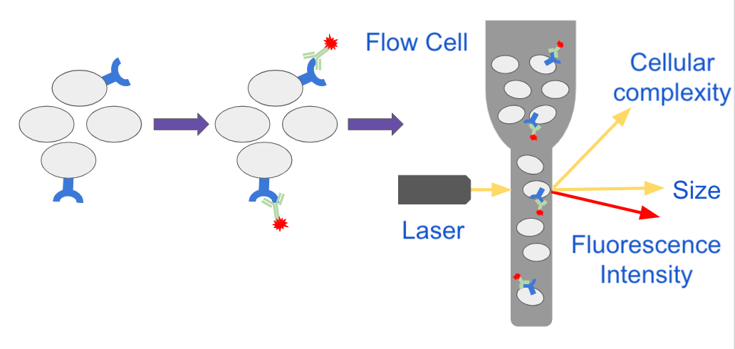
Flow cytometry is a technique that employs an optical-electronic detection apparatus to analyze the physical and chemical properties of microscopic particles.
. In this article we break down the 4 main types of cytometers. The resulting read-out provides information on cell quantity and potential. IFC provides quantitative spatially registered image data for every event analyzed which allows the morphometric.
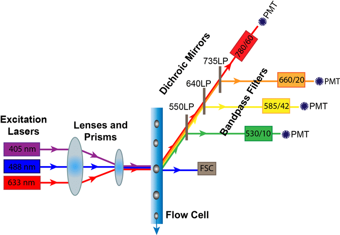
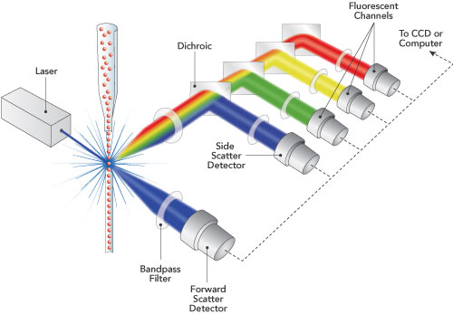
IMAGING FLOW CYTOMETRY Flow cytometry is finding increasing use in clinical laboratories for the diagnosis and monitoring of disease. Request PDF On May 1 2019 Keisuke Goda and others published In Flow Cytometry Image Is Everything Find read and cite all the research you need on ResearchGate. Principles of the Flow Cytometer Flow Cytometry Basics Guide 3 1 Principles of the Flow Cytometer Fluidics System One of the fundamentals of flow cytometry is the ability to measure the properties of individual particles.
Flow cytometry from the greek words cyto cell and metry measure is a powerful technique that can provide us with information about the properties of cells morphology cellular properties cell cycle stage etc. With the combined advantages of optical microscopy and flow cytometry imaging flow cytometry IFC has quickly become an established tool for performing cytometric analysis in diverse areas of biology including microbiology immunology and stem cell biology 1-14IFC provides quantitative spatially registered image data for every event analyzed which allows the morphometric. This information can be used to individually sort or separate subpopulations of cells.
Deep learning With the combined advantages of optical microscopy and flow cytometry imaging flow cytometry IFC has quickly become an established tool for performing. Flow cytometry has become a powerful tool for biological research and clinical diagnostics and its applications have been essential to innu-merable advances in cell biology and immunology as well as for understanding diseases such as im-munodeficiency and cancer56. Imaging flow cytometry has been used to study erythrocytes erythroid cell maturation sickle cell disease.
Imaging Cytometers. 00131105 And at this stage I just want to make a point here 00131418 that at the time of recording this seminar 00131711 the only commercially available imaging flow cytometer 00131919 is from a. With an advent of.
A CYTO 2016 Scientific Tutorial presented by Andrew Filby PhD. 69A1037 1042 Flow Server T drive Flowdata Flow Resources Flow References Isotypes. When a sample enters a flow cytometer the particles are randomly.
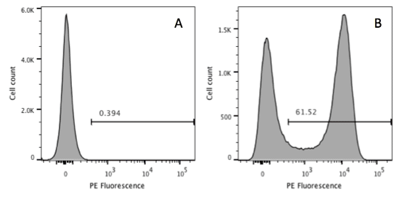
Typically this information is about antigens expressed by these cells either on the cell surface or less commonly in the interior of the cell. Its great advantage lies on the potential to analyze individual cells in a population without averaging in contrast to eg Western blot. Can use isotype control to test how well the blocking buffer worked.
With the combined advantages of optical microscopy and flow cytometry imaging flow cytometry IFC has quickly become an established tool for performing cytometric analysis in diverse areas of biology including microbiology immunology and stem cell biology114. Goda K123 Filby A4 Nitta N12. Its working depends on the light scattering features of the cells under investigation whi.
Thanks for the A2A. Flow Cytometry Flow Cytometry is the technical process that allows for the individual measurements of cell fluorescence and light scattering. Image Cytometry for the Flow Cytometrist.
Flow cytometry controls instrument setup and determination of positivityCytometry Part A 2006. Imaging flow cell sorters and time-lapse. Imaging flow cytometers IFC combine traditional flow cytometry with fluorescence microscopy.
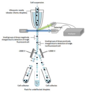
When Image Really is Everything Open to view video. Imaging Cytometer What Is an Imaging Cytometer. This process is performed at rates of thousands of cells per second.
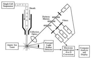
As the name implies imaging cytometers statically image a large number of cells and then process the images. Light is detected by a photomultiplier tube PMT or a photodiode which converts it via a pre-amplifier to a voltage ie an electrical output that is proportional to the original. In Flow Cytometry Image Is Everything.
In Flow Cytometry Image Is Everything Keisuke Goda123 Andrew Filby4 Nao Nitta12 Key terms imaging flow cytometry. Applications of imaging flow cytometry can be used for the assessment of acute leukemia. Epub 2019 May 3.
Flow cytometry is concerned with the measurement of the light intensity of a cell whether it be scattered laser light or fluorescence emitted by a fluorochrome. One of the most common applications is in the diagnosis of leukemia and lymphoma. Flow cytometry is a sophisticated instrument measuring multiple physical characteristics of a single cell such as size and granularity simultaneously as the cell flows in suspension through a measuring device.
This allow for rapid analysis of a sample for morphology and multi-parameter fluorescence at both a single cell and population level Barteneva Fasler-Kan Vorobjev 2012IFC can track protein distributions within individual cells like a confocal or fluorescence. MaeckerHT and Trotter J. Flow cytometry is a laboratory method used to detect identify and count specific cells from blood bone marrow body fluids such as cerebrospinal fluid CSF or tumors.
Image analysis while allowing the same multiparametric capabilities as flow cytometry also permits visualization of the measured events for better quantification of less cellular specimens such as fine needle samplesIntroduction. 2 color viability flow cytometry 96 well plate Guava is 60 min some faster systems are closer to 20 min but are not counting everything in the plate Accumen claims 20 min for counting.
Sensific Gmbh Imaging Flow Cytometry

Representative Flow Cytometric Cytogram And Histogram A Cytogram Of Download Scientific Diagram
Analyzing Flow Cytometry Results Nanocellect

Flow Cytometry Vector Illustration Flow Cytometry Vector Illustration Illustration

Flow Cytometery Mybiosource Learning Center
Flow Cytometry Archives Bitesize Bio
Chapter 4 Data Analysis Flow Cytometry A Basic Introduction
Analyzing Single Cells With Flow Cytometry

Flow Cytometry Analysis Of Intracellular Uptake Of Fitc Hsa Nps Gated Download Scientific Diagram

View Of An Adapted Novel Flow Cytometry Methodology To Delineate Types Of Cell Death In Airway Epithelial Cells Journal Of Biological Methods

Diagnostic Potential Of Imaging Flow Cytometry Trends In Biotechnology

Flow Cytometry Antibodies Tips Flow Cytometry Flow Life Science

Flow Cytometry And The Sheath Fluid You Use Lab Manager
How Does Flow Cytometry Work Nanocellect
How Does Flow Cytometry Work Nanocellect
Putting Down A Marker In Flow Cytometry To Help Determine Positivity

How To Identify Bad Flow Cytometry Data Bad Data Part 1 Cytometry And Antibody Technology

Resolution Of Flow Cytometry Data What It Means And Why It Matters For Publications Marissa Fahlberg Phd
Using Application Settings To Standardize Flow Cytometry Results Across Experiments And Instruments Sanguine Bio Researcher Blog


0 komentar:
Posting Komentar